
FIG. 1.
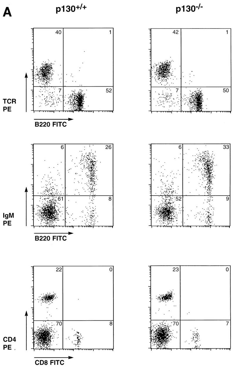

Normal differentiation and proliferation of p130−/− lymphocytes. (A) Wild-type and p130−/− splenocytes were stained for expression of the indicated cell surface markers and analyzed by flow cytometry. The percentages of cells in each quadrant are indicated. Each plot displays 2,000 events. FITC, fluorescein isothiocyanate; IgM, immunoglobulin M. (B, inset) Background incorporation of [3H]thymidine in cultures of unstimulated wild-type (open bar) and p130−/− (solid bar) lymph node lymphocytes cultured for 2 h in the presence of 1 μCi of [3H]thymidine. (B and C) Time course of [3H]thymidine incorporation, as a measure of DNA synthesis, upon PMA-plus-ionomycin (B) or concanavalin A (con A) (C) stimulation of lymph node lymphocytes (see Materials and Methods). Triplicate cultures were pulsed for 2 h at the times shown. Open squares, wild type; closed circles, p130−/−. Results are presented as mean counts per minute ± standard errors of the mean. Data are representative of three independent experiments. Similar results were obtained with wild-type and p130−/− purified splenic T cells. (D) In vivo T-cell responses as determined by contact hypersensitivity. The inflammatory responses of p130+/+ and p130−/− animals in reaction to topical application of exogenous hapten (2% oxazolone in ethanol) (solid bars) are presented as change in ear thickness 48 h after challenge. The control (open bars) represents the response to ethanol without hapten. Naive animals were not sensitized prior to challenge. Error bars indicate standard errors of the means.