
FIG. 2.
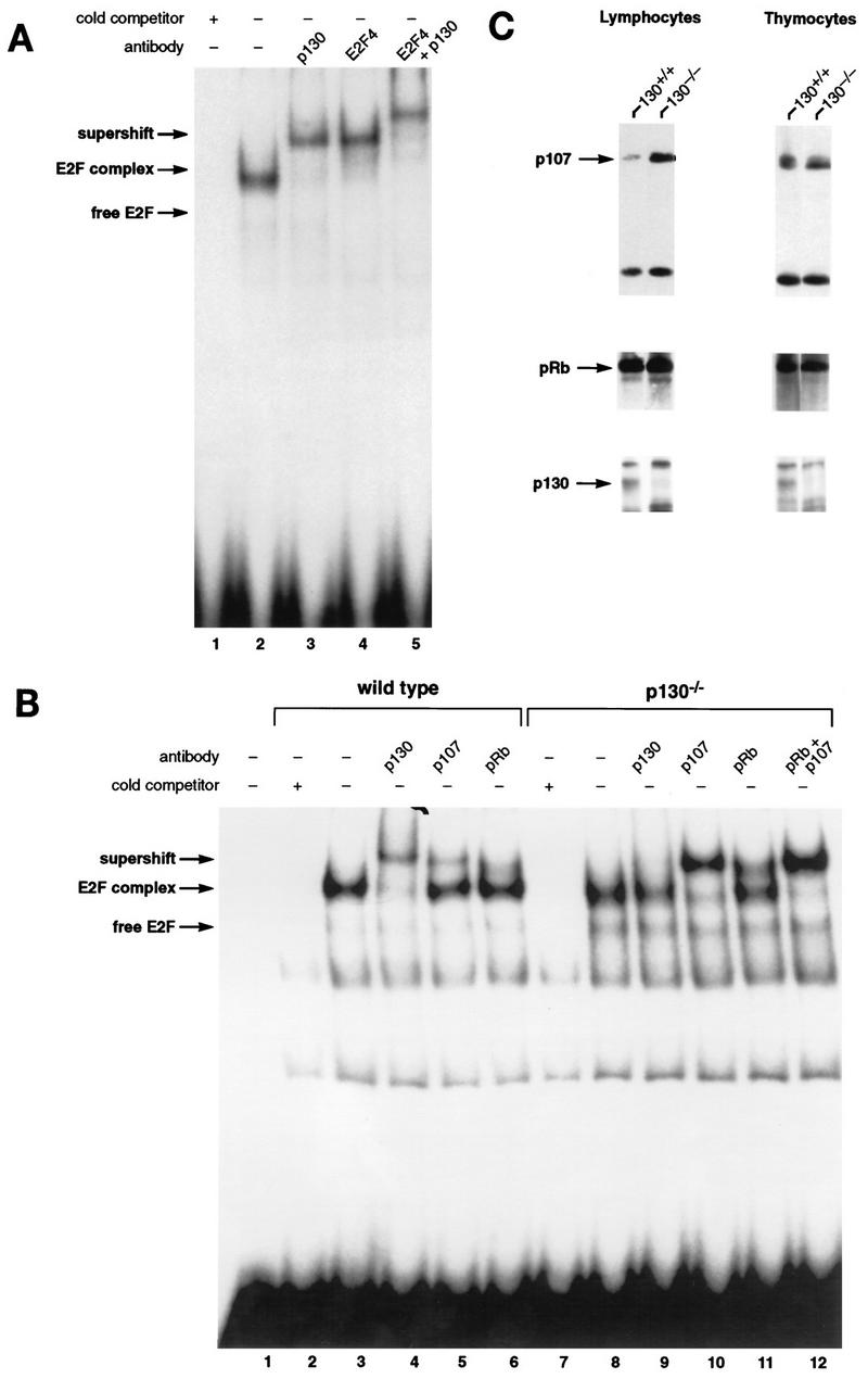
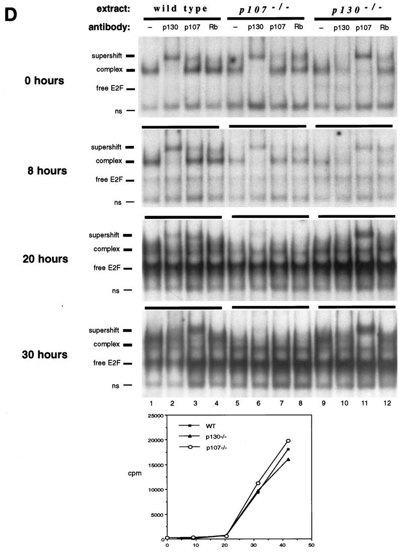
Characterization of E2F complexes in wild-type and mutant peripheral T lymphocytes. (A) Whole-cell extracts of wild-type lymph node lymphocytes were analyzed by EMSA with a radiolabeled E2F probe. The single complex (lane 2) was supershifted by antibody to p130 (lane 3) and antibody to E2F-4 (lane 4). Addition of both antibodies induced a second supershift of the entire complex (lane 5). Specificity of the complex was demonstrated by competition with a 100-fold excess of cold E2F oligonucleotide (lane 1). (B) Characterization of E2F complexes present in wild-type and p130−/− T-lymphocyte extracts. The complex present in p130−/− cells (lane 8) appears to be similar in mobility to that in wild-type cells (lane 3). These complexes were analyzed with antibodies to p130 (lanes 4 and 9), p107 (lanes 5 and 10), or pRB (lanes 6 and 11). The positions of free E2F and higher-order E2F complexes are indicated. The specificity of these complexes was shown by competition with a 100-fold excess of cold E2F oligonucleotide (lanes 2 and 7). Analyses of purified T-cell populations or lymphocyte nuclear extracts yielded similar results (data not shown). (C) Elevated expression of p107 in peripheral T lymphocytes of p130−/− mice. Whole-cell extracts (30 μg) from wild-type and p130−/− thymocytes and lymph node T lymphocytes were subjected to immunoblot analysis with p107, pRB, or p130 antibodies. The lower band in the p107 panel is a cross-reacting protein that serves as a loading control. Similar results were obtained upon analyses of nuclear extracts. Quantitation indicates that p107 levels increased 25% in thymocytes and sevenfold in peripheral lymphocytes. (D) E2F complex analysis as wild-type and mutant T cells reenter the cell cycle. Lymph node lymphocytes were isolated from wild-type (lanes 1 to 4), p107−/− (lanes 5 to 8), and p130−/− (lanes 9 to 12) animals and stimulated with PMA-ionomycin. Cell extracts were prepared at the indicated time points, and E2F complexes were identified by using antibodies to p130, p107, and pRB. In addition, a fraction of each culture was labeled with [3H]thymidine to determine S-phase entry (bottom panel). The positions of the nonspecific DNA binding complex (ns), free E2F, E2F complex, and antibody-induced supershift are indicated at the left.