FIG. 5.

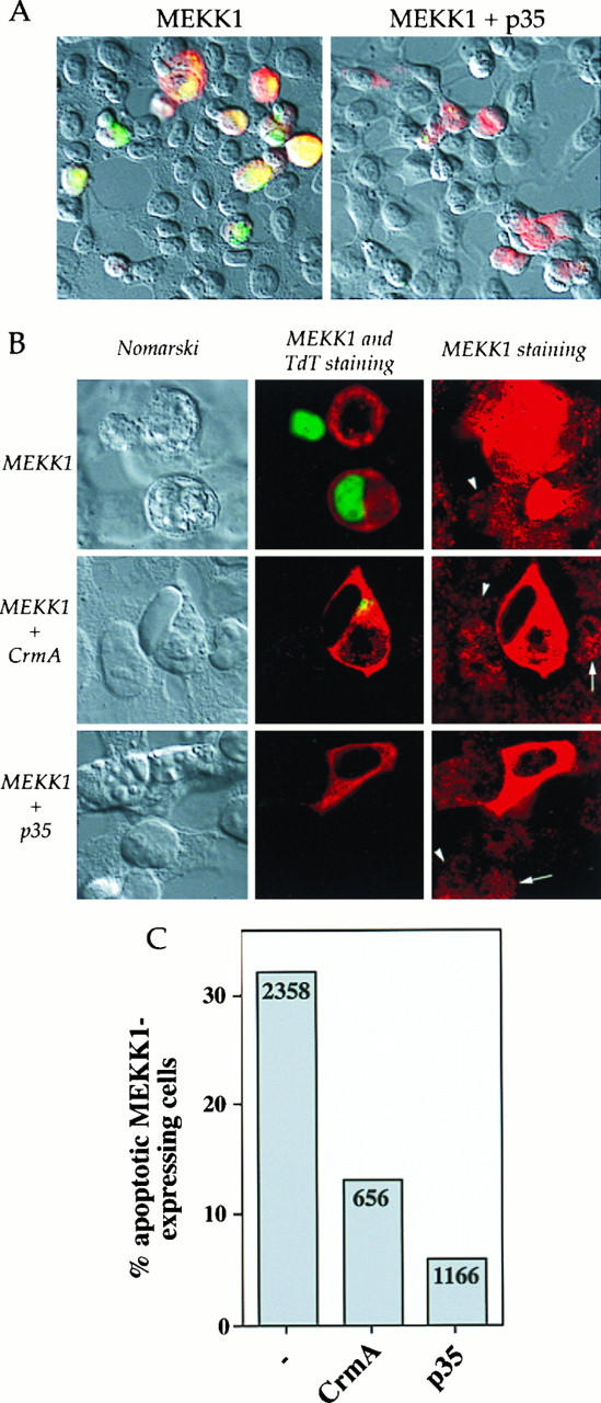
p35 and CrmA inhibit MEKK1-induced DNA fragmentation in HEK293 cells. Cells were transfected with 0.5 μg of MEKK1.cp4 alone or in combination with 2 μg of either p35.cp_ or CrmA.cp_. Two days later, the cells were stained for MEKK1 expression and for DNA fragmentation. (A) Nomarski views (magnification, ×40) of HEK293 cells transfected with MEKK1 or with MEKK1 and p35 and overlaid with fluorescent staining for MEKK1 expression (red staining) and for DNA fragmentation (green staining). (B) Views (magnification, ×160) of HEK293 cells transfected with MEKK1 alone or in combination with either CrmA or p35. (Left panels) Nomarski views. (Middle panels) Fluorescent staining for MEKK1 expression (red staining) and DNA fragmentation (green staining). (Right panels) Fluorescent staining for MEKK1 expression. In these last views, an exposure longer than that in the middle panels was used to visualize the localization of endogenous human MEKK1. The arrows indicate granular cytoplasmic localization, while the arrowheads indicate nuclear localization. (C) Quantitation of the percentage of MEKK1-transfected cells, in the presence or in the absence of the indicated proteins, that showed DNA fragmentation. The numbers in the columns indicate the number of cells transfected with MEKK1 and counted on at least four coverslips from at least two different experiments.
