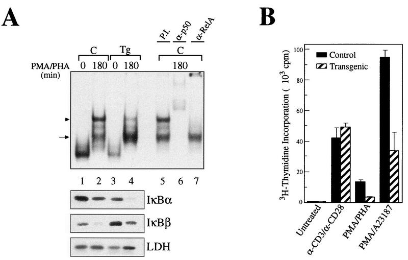
FIG. 6.
Splenic T cells from transgenic animals show impaired proliferation in response to some mitogenic signals. (A) EMSA with a palindromic κB and nuclear extracts from control (C, lanes 1 and 2) or transgenic (Tg, lanes 3 and 4) splenic T cells treated with PMA-PHA for 180 min. Preincubations with preimmune serum (P.I., lane 5) or with the respective antibodies (lanes 6 and 7) allowed the identification of the complexes as p50/RelA heterodimers (arrowhead) and p50-containing complexes, possibly homodimers (arrow). The corresponding cytoplasmic extracts were analyzed by Western blotting with antibodies against IκBα, IκBβ, and LDH. (B) Splenic T cells were stimulated with anti-CD3 plus anti-CD28, PMA-PHA, or PMA-A23187 over a period of 3 days in the presence of 0.5 μCi of [3H]thymidine per 100 μl. Each column represent the results of six independent measurements.

