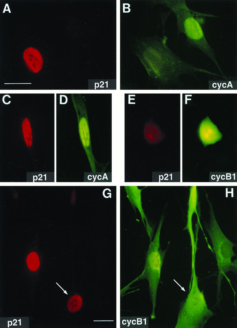
FIG. 1.
Nuclear colocalization of p21 with cyclin A and cyclin B1 in exponentially growing normal human fibroblasts. Asynchronous normal HDF (Hs68) were fixed in paraformaldehyde and simultaneously stained with mouse monoclonal anti-p21 (red; Texas red) and with rabbit polyclonal anti-cyclin A or anti-cyclin B1 (green; fluorescein) antibodies as described in Materials and Methods. Representative micrographs of cells in G1 phase (A and B), S phase (A, B, G, and H), and G2 phase (C, D, G, and H), and mitosis (E and F) are shown. Cyclin A accumulates in the nucleus in the beginning of the S phase, whereas cyclin B1 accumulates during late S phase and in G2 in the cytoplasm and enters the nucleus at the onset of mitosis (27). A cell with both cytoplasmic and nuclear cyclin B1 accumulation is marked with an arrow (G and H). Quantitation of localization experiments is shown in Tables 1 and 2. Exposure times for given antigen were constant for all micrographs. Bars, 10 μm.

