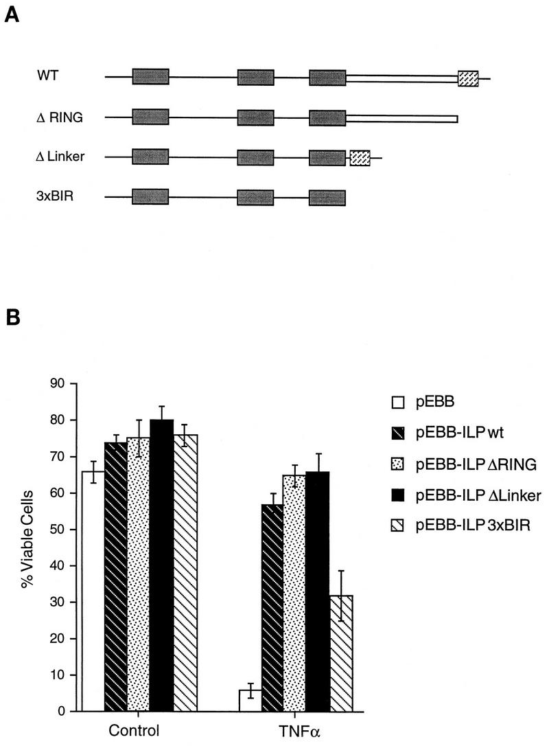
FIG. 2.
Deletion analysis of hILP. (A) Schematic diagram of hILP showing deletion mutants. BIR domains (solid boxes), the amphipathic region (open box), and the ring finger (hatched box) are shown. WT, wild type. (B) MCF7F cells were transfected with pCMV-lacZ and either pEBB, pEBB-hILP, or pEBB deletion mutants of hILP. After 24 h, medium was removed and replaced with either fresh medium alone or fresh medium plus rhTNF-α. Cells were incubated for 12 h, at which time cells were fixed, stained for β-galactosidase expression, and scored for apoptotic morphology. The expression of FLAG-tagged deletion constructs was confirmed by immunoblotting with anti-FLAG bioM5 (Kodak) (data not shown). Results are expressed as percent viable cells (number of flat blue cells/number of flat and round blue cells × 100). The data represent the means ± standard deviations (n = 3).

