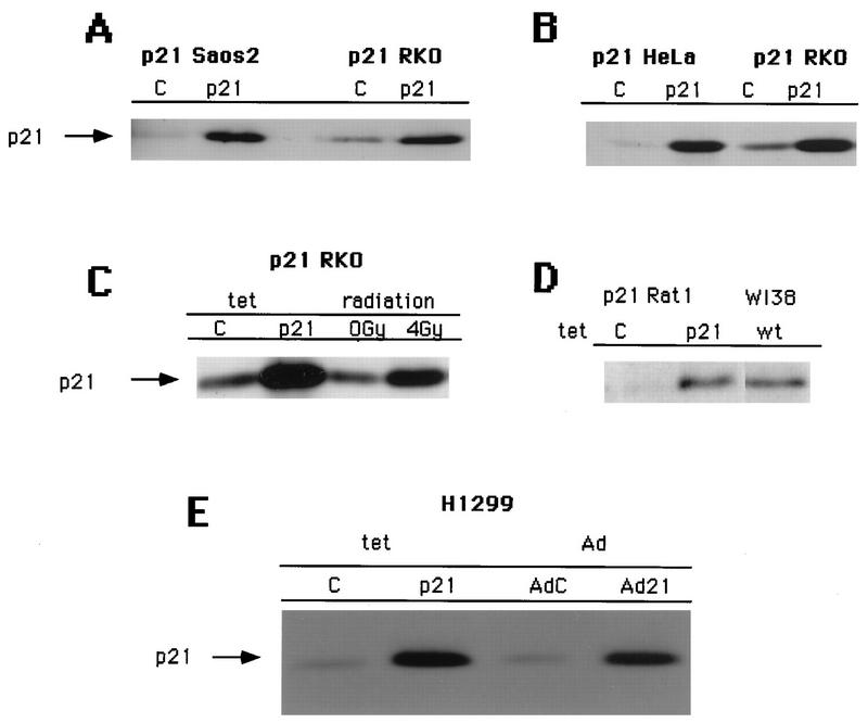
FIG. 1.
Tetracycline-controlled expression of p21 in a panel of pRb-positive and pRb-negative cell lines. Shown are Western blots of total-cell lysates (0.2 mg of protein) with an antibody against p21 (C-19). Tetracycline-controlled p21 expression in uninduced cells (lanes C) and induced cells (lanes p21) is demonstrated. Equal numbers of cells were plated at low density (see Materials and Methods) and incubated for 3 days in medium with or without tetracycline. (A) Comparison of the levels of expression in Saos2 Tet p21 and RKO Tet p21 cells. (B) Comparison of the levels of expression in HeLa Tet p21 cells and RKO Tet p21 cells. (C) Comparison of the levels of expression in RKO Tet p21 cells and in RKO cells subjected to 0 or 4 Gy of ionizing radiation. (D) Comparison of the levels of expression in Rat1 Tet p21 cells and the levels in WI38 normal human diploid fibroblasts approaching senescence. (E) Comparison of the levels of expression in H1299 Tet p21 and adenovirus (Ad)-transduced H1299 cells (with an MOI of 200).

