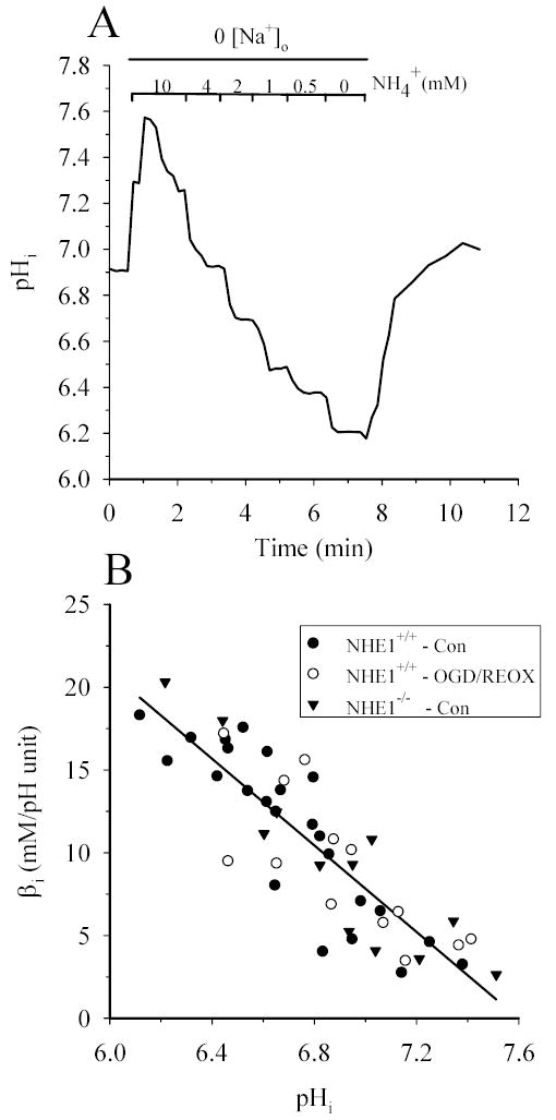
Figure 1. Intrinsic buffer capacities (βi) in NHE1−/− and NHE1+/+ astrocytes are similar and unchanged following OGD/REOX.

Intrinsic buffer capacity was determined in NHE1−/− and NHE1+/+ astrocytes and in NHE1+/+ astrocytes following 2 h OGD and 1 h REOX. Cells were sequentially exposed to decreasing concentration of NH4+ in Na+-free HEPES-MEM and the change in pHi determined. A. A representative trace of pHi in a single NHE1+/+ cell in response to changes in NH4+. B. βi was plotted as a function of pHi in normoxic NHE+/+ astrocytes, NHE1−/− astrocytes, and NHE1+/+ astrocytes following OGD/REOX (three cultures). There was no significant difference in the regression fits among these groups (p > 0.05).
