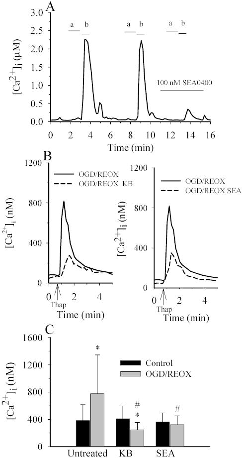
Figure 9. NCX contributes to an increase in [Ca2+]i in astrocytes following OGD/REOX.

A. Reverse-mode of NCX was induced by exposing cells with Ca2+-free HEPES buffer containing gramicidin (2 mg/ml) for one min (a), and subsequently to a Na+-free buffer (1.2 mM Ca2+) for 30–40 sec (b). Cells were then returned to normal HEPES-MEM. The increases in [Ca2+]i via the reverse-mode of NCX were reproducible. However, it was decreased by ~90% in the presence of 0.1 μM SEA0400. B. Thapsigargin-induced Ca2+ release was determined under control (1.2 mM) [Ca2+]o conditions in NHE1+/+ astrocytes following 3 h normoxia (CON) or 2 h OGD plus 1 h REOX (OGD/REOX). In some OGD/REOX studies, cells were exposed to 3 μM KB-R7943 or 0.1 μM SEA0400 throughout the OGD/REOX and Ca2+ release experiment. Data points are the average of 20 cells from a representative experiment. C. Summary of peak thapsigargin-induced Ca2+ release data for CON or OGD/REOX with and without either 3 μM KB-R7943 or 0.1 μM SEA0400. Data are means ± SD (n = 80–120 cells, 3 cultures). * p < 0.001 vs. baseline control; # p < 0.001 vs. OGD/REOX.
