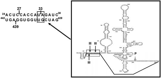
Figure 8.
Diagram including the proposed structural motif which contains both RNase III cleavage domains and encloses the HCV IRES. On the left is shown the interaction between nt 24 and 38 of the 5′ (UTR) with the nt 428–442 of the HCV Core–coding sequence. The arrows represent the RNase III cleavage sites. The ‘loop’ is shown within a box on the right of the figure. Symbol ‘P’ refers to the cleavage position of human RNase P, which is another RNA structure dependent nuclease. After the annealing, both RNase III and RNase P cleavage sites became in close proximity.

