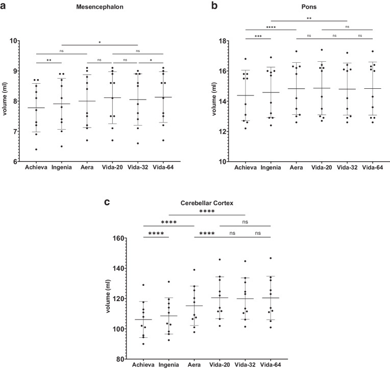
Fig. 7.
Results of post-hoc analysis between the different MRI-scanners. Vida-20, Vida-32 Fig. 2: Results of post-hoc analysis between the different MRI-scanners for a the mesencephalon, b the pons, c the cerebellar cortex. Vida-20, Vida-32 and Vida-64 represent the results of Siemens MAGNETOM Vida 3T with the 20-, 32- or 64-channel receiver coil respectively. ns not significant. *: p < 0.05; **: p < 0.005; ***: p < 0.0005; ****: p < 0.0001

