
Figure 2.
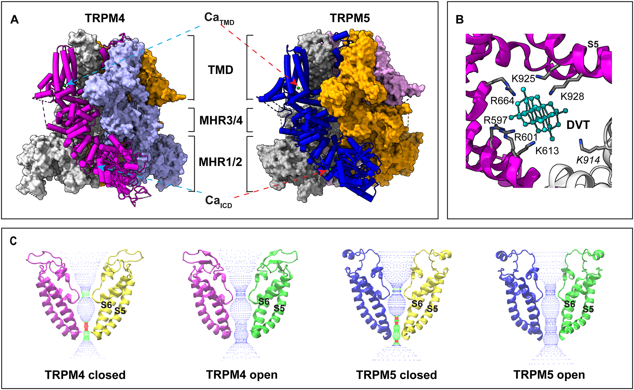
Structural features of TRPM4 and TRPM5 revealed by cryo-EM. A. Overall structure of TRPM4 (PDB ID 9B8Y, left panel) and TRPM5 (7MBQ, right panel). The light green spheres represent bound Ca2+ ions. TMD, transmembrane domain; ICD, intracellular domain; MHR, melastatin homology region. B. The binding pocket of decavanadate (DVT, cyan) in TRPM4 (9B8Y). Two neighboring subunits are colored in magenta and gray, respectively. Key positively charged residues in direct contact with DVT are highlighted. C. The closed states and open states of TRPM4 and TRPM5 (PDB ID from left to right: 6BQR, 9B8Y, 7MBR, and 7MBQ), highlighting the ion permeation pore. S5 and S6 represent the fifth and sixth transmembrane helices, respectively.