
Figure 3.
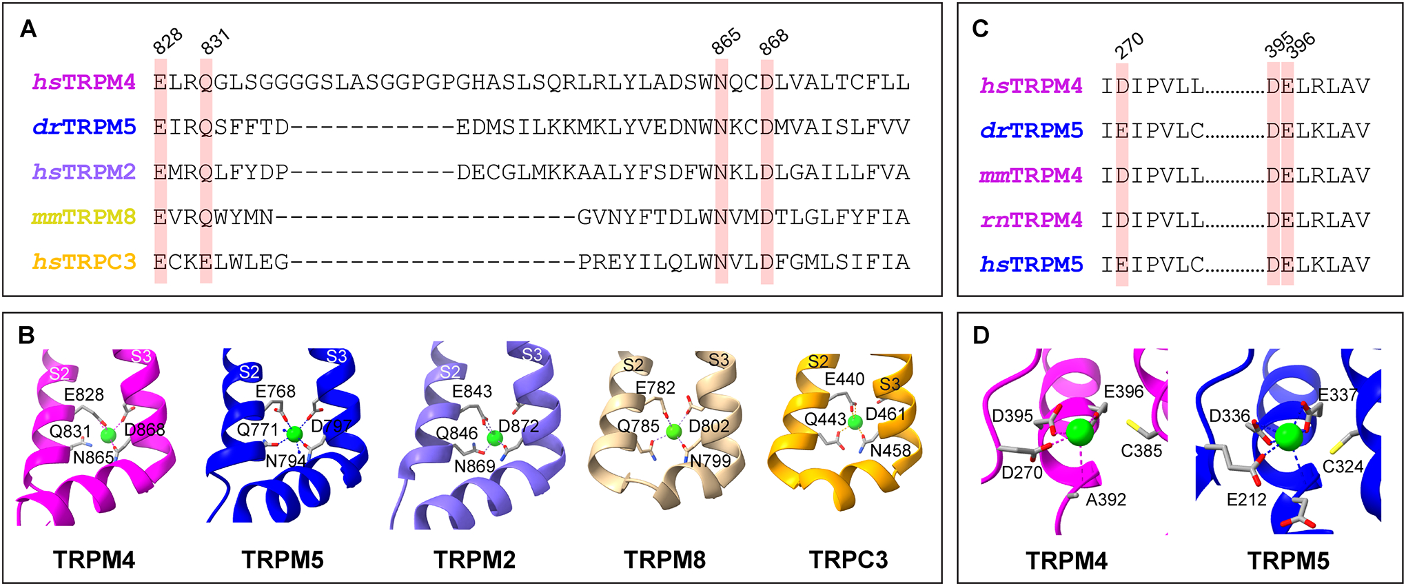
Calcium binding sites of representative TRP channels. A. Amino acid sequence alignment of the S2-S3 regions of human (hs) TRPM4, zebrafish (dr) TRPM5, human TRPM2, mouse (mm) TRPM8, and human TRPC3, with the key conserved residues for calcium binding highlighted. B. Transmembrane calcium binding pockets of hsTRPM4 (magenta, 9B8Y), drTRPM5 (deep blue, 7MBQ), hsTRPM2 (purple, 6PUS), mmTRPM8 (light yellow, 7WRB), and hsTRPC3 (golden, 7DXB). S2 and S3 represent the second and third transmembrane helices, respectively. C. Amino acid sequence alignment of the intracellular N-terminal calcium binding sites of TRPM4 and TRPM5 of several species, with the key conserved residues highlighted. D. Intracellular calcium binding pockets of TRPM4 (magenta, 9B8Y) and TRPM5 (deep blue, 7MBQ). The light green spheres represent bound Ca2+ ions. Amino acid numbers are for human TRPM4.