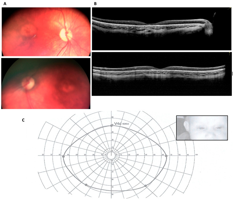
Figure 1.
Bilateral bull’s eye maculopathy (BEM) as detected at fundus examination and OCT scans performed in the corresponding region at 7 months. (A). Bilateral BEM is characterized by a round hypopigmented central zone surrounded by a hyperpigmented ring. Top image: right eye (RE); bottom image: left eye (LE). (B). OCT images of the macula. A very thin fovea (total foveal thickness of 72 µm in RE and 78 µm in LE) is visible in both eyes due to the development of foveal atrophy involving only the outer retinal layers. Top image: RE, oblique scan; bottom image: LE, vertical scan. (C). The binocular attraction visual field shows a normal extension for the age. The infrared image of the child’s face during the examination is shown in the top right window and allows us to identify the fixation changes made by both eyes and the head.

