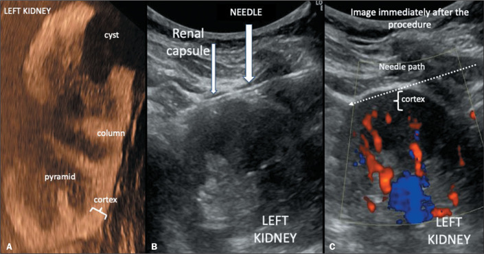
Figure 1.
A: Arterial-phase ultrasound image with microbubble contrast showing hyperenhancement of the cortex and hypoenhancement of the renal pyramids, which are poorly defined in the normal kidney. The cortex above the pyramids is only 5 mm thick. B: B-mode ultrasound image of a cortical tangential biopsy of the left kidney: the needle passes just below the renal capsule, collecting material only from the renal cortex, avoiding the interlobar arteries, which are visible on color Doppler (C) mapping (scale limit: 11 cm/s) and appear in red in the image on the right, taken immediately after the needle was removed, in which no blood reflux is observed along the needle path (dotted arrow). In this image, it is clear that the subcapsular cortical tangential path does not reach the interlobar or arcuate arteries. Care should be taken to avoid a puncture in the region close to cysts, because they can deform the cortex and change the position of the arteries.

