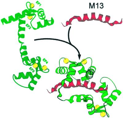
Figure 1.
Decoding of the Ca2 + signal by conformational changes in EF hand proteins (CaM). CaM interacts with a 26-residue binding domain (red peptide, top right) of a skeletal muscle myosin light chain kinase termed M13. CaM (left) has bound Ca2+ (yellow shares) to its four EF hands. It has already undergone the change that has made its surface more hydrophobic, but it still is in the fully extended conformation. The interaction with M13 collapses it to a hairpin shape that engulfs the binding peptide.

