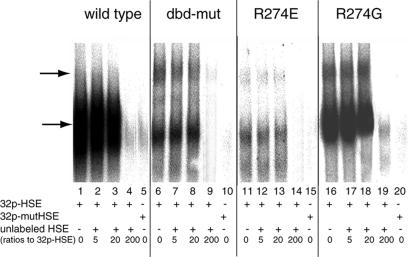
Figure 2.
DNA-binding assay. Gel mobility shift assay of wild-type and mutant DBDs to labeled HSE is shown. A mutant HSE with nGAAnnTTCn repeats changed to nGAAnnGGCn was also used to ensure binding specificity (lanes 5, 10, 15, and 20). Increasing amounts of unlabeled HSE were added for competition assay. The numbers shown at the bottom of each lane are the ratios of unlabeled HSE to [32P]HSE. Major DBD–HSE complexes are shown by the lower arrow. Mutant DBDs also form higher-order complexes (upper arrow). Note that microgram quantities of HSFs are used in these experiments because the recombinant proteins bind to the HSE less well than the native HSF.

