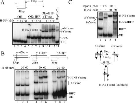
Figure 3.
Electrophoretic band-shift assays with H-NS. (A) H-NS band-shift with OE substrate containing 49 bp of flanking donor DNA. (H-NS-t'some) H-NS-bound transpososome; (uf-t'some) unfolded transpososome; (f-t'some) folded transpososome; (IHFC) IHF complex; (OE) outside end DNA. In lane 13, heparin and CaCl2 were added after transpososome formation to unfold the transpososome. (B) H-NS band-shift with transpososomes formed with OE DNA containing different lengths of flanking donor DNA. Note that H-NS also failed to bind a transpososome formed with no flanking donor DNA (data not shown). (C) Effect of H-NS on transpososome mobility when H-NS is added to folded and unfolded transpososomes. As depicted, heparin and CaCl2 were added to unfold the transpososome prior to H-NS addition; based on data from Figure 4, H-NS (hexagon) is shown bound to the flanking donor DNA. Note that in C, OE DNA, IHF, and transposase were present at five, three-, and threefold lower concentrations, respectively, relative to A and B.

