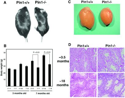
Figure 1.
Reduced body weight and testicular atrophy in Pin1−/− mice. (A and B) Reduced body weight. Representative adult wild-type mouse (Left) and Pin1−/− mouse (Right) are shown in A. A comparison of body weight of 10 wild-type and Pin1−/− mutant male and female mice at ≈3.5 and ≈7 months is shown in B. (C and D) Testicular atrophy, as indicated by representative testis from wild-type or Pin1−/− mouse at ≈3.5 months old (C) or by histopathological comparison (D). Testicular sections obtained from ≈3.5- and ≈16-month-old mice were stained with hematoxylin and eosin.

