Figure 3.
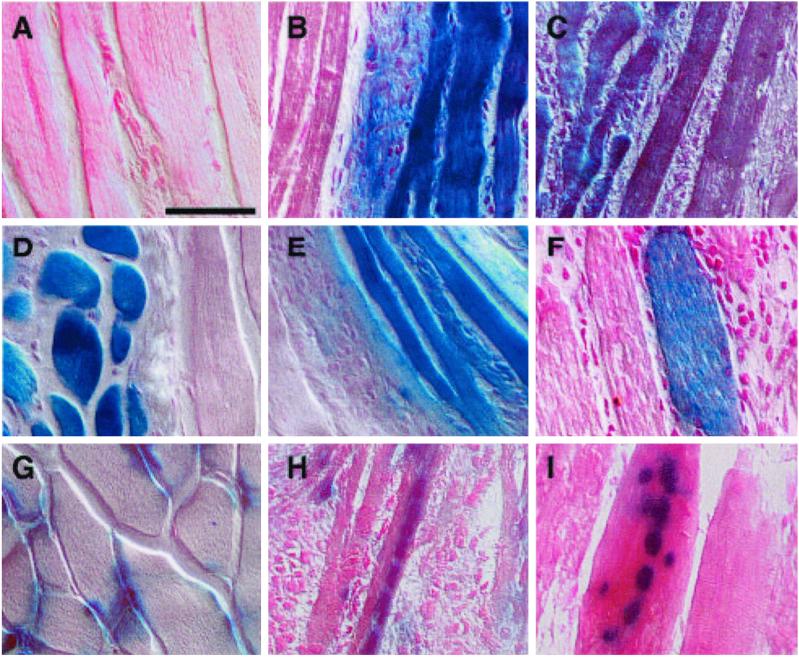
In vivo myogenic potential of muscle-derived cells. Muscle-derived cells were purified from C57BL/6-ROSA26 (B–F) or C57/MlacZ (H, I) mice by flow cytometry or magnetically followed by flow cytometry. Test populations were injected into the TA muscles of non-ROSA26 animals that had been injected with cardiotoxin 24 h earlier. The TA muscles were removed from recipients 2–3 weeks later, cryosectioned, and stained with X-Gal for β-galactosidase expression. (Bar = 50 μM.) (A) Injured muscle injected with HBSS. (B) Muscle injected with unfractionated muscle-derived cells. The large blue tracts are fibers regenerated from Rosa26-derived cells. (C) Incorporation of Sca-1-positive muscle cells. (D) Incorporation of Sca-1-negative muscle cells. (E) Incorporation of Sca-1+/CD45+ cells. (F) Incorporation of Sca-1+/CD45− cells. (G) Section of unmanipulated C57/MlacZ muscle. Note the nuclear localization of lacZ at the edges of the fibers shown in cross section. (H) CD45-positive C57/MlacZ-derived cells injected into regenerating muscle and sectioned longitudinally. (I) CD45-negative C57/MlacZ incorporation.

