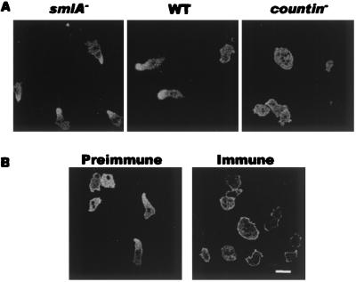
Figure 5.
CF regulates the distribution of GFP–myosin in developing cells. Cells expressing GFP–myosin were starved in a chambered cover glass slide. (A) The cells were mixed 1:9 with the indicated cell line. (B) The cells were mixed with wild-type cells, and preimmune or immune anti-countin antibodies were added 1 h after starvation. The distribution of GFP–myosin in the live cells was recorded 5 h later. (Bar is 10 μm.)

