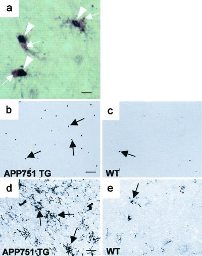
Figure 3.
APP751 TG mice show increased expression of inflammatory markers after 24-h MCA occlusion. (a) Double immunohistochemical staining with phospho-p38 MAPK ab (Ni-diaminobenzidine, purple) and F4/80 ab (diaminobenzidine, brown). p38 MAPK activity (arrowheads) is localized to the nuclei of F4/80 positive microglial cells (arrows). (b) p38 MAPK activity (arrows) in peri-infarct region of an 8-month-old APP751 TG mouse demonstrated by phospho-p38 MAPK immunohistochemistry. (c) An age-matched WT mouse shows only few cells with p38 MAPK activity (arrows) in perifocal cortex. (d) F4/80 ab detects robust microgliosis (arrows) in the perifocal cortex of an APP751 mouse. (e) Immunoreactivity for F4/80 is much lower in the perifocal cortex of a WT mouse. [Bar = 8 μm (a), 100 μm (b and c), and 50 μm (d and e)].

