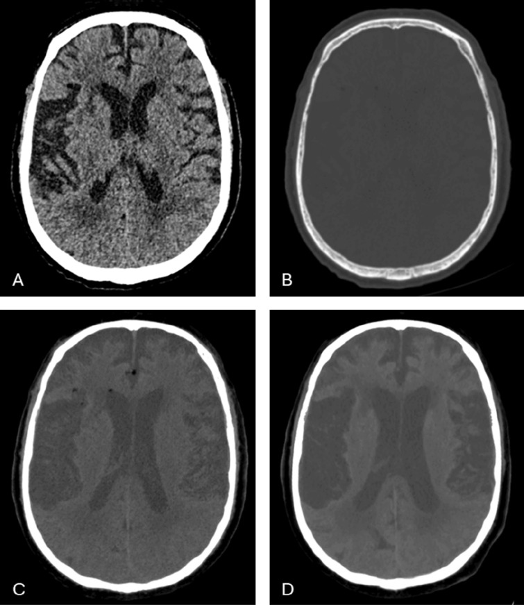
Figure 2.
The subarachnoid and intraventricular fat droplets are hardly visible on standard brain window (A) and very subtle on bone window (B). 10 mm minimum intensity projection (MinIP) images in the soft tissue window show the true extend of the fat dissemination in the cerebrospinal fluid (C). These were not present on the initial post‑traumatic NCCT (D).

