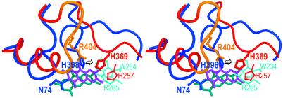
Figure 6.
Comparison of the α-subunit of APS reductase with fumarate reductase (stereo view). The structural alignment of the α-subunit of APS reductase and the flavoprotein subunit of fumarate reductase from W. succinogenes is shown. For APS reductase, the residues of the FAD-binding domain are shown in blue, the residues of the capping domain are shown in cyan and the FAD in blue-green. For fumarate reductase, the residues of the FAD-binding domain are shown in red and the insert in orange, and the residue of the capping domain is shown in light red and the FAD in magenta.

