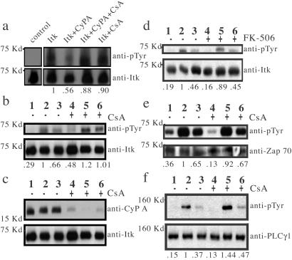
Figure 4.
(a) CypA, CypA/CsA complex, or CsA was added to baculovirus-expressed Itk immediately before resuspension in ATP/kinase buffer. A mock kinase assay of Itk (left lane) was performed in which ATP was excluded from the kinase reaction buffer. Itk phosphorylation is normalized to Itk protein levels, and net changes in phosphorylation are indicated below each lane. (b–f) Lanes 1, 2, and 3 are mock stimulation, 40 s and 2 min, respectively, following TCR stimulation of Jurkat cells in the absence (−) of drug treatment, whereas lanes 4, 5, and 6 represent the same conditions in the presence (+) of drug. (b) Itk phosphorylation following TCR stimulation of Jurkat cells in the presence and absence of CsA. (c) CypA binding to Itk was monitored by detection of Itk immunoprecipitates with an anti-CypA antibody. (d) Itk phosphorylation in the presence and absence of FK-506. (e) Zap-70 phosphorylation and (f) PLCγ1 phosphorylation in the presence and absence of CsA. Phosphorylation levels of Itk, Zap-70, and PLCγ1 are normalized to total protein in each lane. Net changes in phosphorylation relative to phosphorylation levels at 40 s following stimulation in the absence of drug (lane 2) are indicated below each lane.

