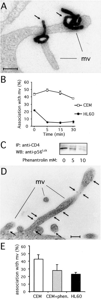
Figure 2.
p56Lck binding to CD4 triggers CD4 anchoring to microvilli. (A) Electron micrograph showing CD4 radiolabeling on microvilli in CEM cells. (Bar = 0.2 μm.) (B) Kinetics of radiolabeled CD4 association with microvilli in CEM and HL60 cells. Data are means ± SE from two to three separate experiments totaling 900–1,200 autoradiographic grains for each time point. (C) p56Lck association with CD4 in CEM cells treated with 1,10-O-phenanthroline for 30 min at 37°C before cell lysis. Data are representative of three independent experiments. (D) Electron micrograph showing CD4 gold labeling on microvilli in CEM cells. (Bar = 0.2 μm.) (E) Quantitation of gold-labeled CD4 association with microvilli at 4°C in CEM cells treated or not treated with 10 mM 1,10-O-phenanthroline and HL60 cells. Data are means ± SE from three to four separate experiments totaling 78 cells per 2,483 gold particles, 40 cells per 1,119 gold particles, and 81 cells per 1,929 gold particles counted for CEM cells, CEM cells treated with 1,10-O-phenanthroline, and HL60 cells, respectively.

