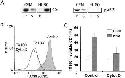
Figure 3.
Disruption of the cytoskeleton increases CD4 TX100 solubility in p56Lck-expressing cells. (A) Western analysis of CD4 and p56Lck solubility by TX100. Cells were lysed in buffer containing 1% TX100 for 20 min at 4°C and then centrifuged at 100,000 × g for 1 h at 4°C to fractionate the lysates in a TX100-soluble (S) and TX100-insoluble (P) fraction. Data are representative of three independent experiments. (B) Typical FACS profile of CD4-associated fluorescence in CEM cells ± 1% TX100 and cytochalasin D (Cyto. D). (C) Quantitation of CD4 solubility by TX100 in CEM and HL60 cells treated or not treated with Cyto. D. Data are expressed as the ratio of fluorescence associated with cells after TX100 addition to fluorescence associated with cells before TX100 addition. Results are means ± SE from three to four independent experiments.

