Figure 3.
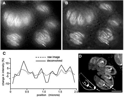
WFM imaging of T. gondii expressing YFP-α-tubulin. (A) Raw WFM YFP fluorescence image of several living parasites with the focal plane near the plasma membrane. (B) Same as A, after processing of the 3D stack by constrained iterative deconvolution. Microtubules are clearly visible as bright striations. Arrows in A and B point to a single microtubule. Compare these with LSCM images in Fig. 1. (C) Plot of the change in intensity along the dashed line in B and the corresponding pixels in A. (D) Cartoon (Lower left) and deconvolved image showing a single focal plane through the interior of a group of parasites revealing conoid, spindle pole, and centriole. Developing daughters are revealed by the U-shaped outline of their growing subpellicular microtubules, capped by the bright conoid. The cartoon of the lowermost parasite in the image shows the substructures within the parasite (see also Fig. 1B). Arrows indicate conoids of mother and daughter parasites. The subpellicular microtubules of the mother (bright perimeter originating at the conoid) extend half the length of the parasite. The distal portion of the cartoon parasite is outlined by the dotted lines. Distal to the daughter conoid, the two dots indicate the location of the centriole and spindle pole, in that order. (Scale bar in A, B, and D, 3 μm.)

