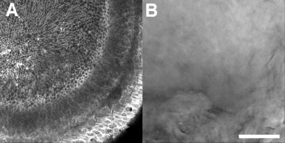
Figure 4.
Comparison of LSCM and WFM performance in imaging of a thick tissue. A 5-day-old quail embryo fixed in formaldehyde and stained with Alexa488-phalloidin is shown. See Supporting Methods for more details. (A) A single LSCM optical section ≈50 μm below the exterior surface of the embryonic eye. Concentrations of actin at cell cortices are visible. (B) The same location in the embryo imaged by WFM recorded with a CCD camera. (Scale bar, 50 μm.)

