
Figure 2.
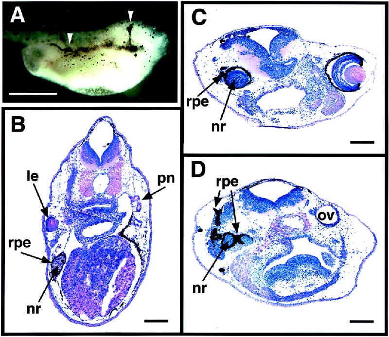
Ectopic eye structures formed in embryos injected with eyeless RNA at the 2-cell stage. Xenopus embryos were injected with various amounts of eyeless RNA into one of the two blastomeres at the 2-cell stage and fixed at stage 41 to 42. Bars indicate 1 mm in A and 0.1 mm in B–D. (A) Embryo injected with 2 ng of ey RNA. Arrows indicate the extension of the RPE toward the midline and along the body trunk. (B) Cross section of an embryo injected with 0.25 ng of ey RNA, showing the ectopic formation of a lens, RPE, and neural retina in the trunk region at the pronephros level. (C) cross section of an embryo injected with 0.5 ng of ey RNA, showing a developmental defect in the region of the original eye with the lack of a lens, the ectopic formation of RPE, and the hyperproliferation in the neural retina. (D) More posterior cross section of the same embryo as in C, showing the ectopic formation of RPE and neural retina in the region of the original otic vesicle. le, lens; nr, neural retina; ov, otic vesicle; pn, pronephros.