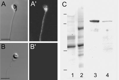
Figure 8.
Detection of the Na+/Ca2+ exchanger in whole sperm and in extracted sperm proteins. (A and A′) Live sperm that were labeled with anti-Na+/Ca2+ exchange IgG exhibited label over the entire sperm with the most intense signal over the midpiece region. (Bar = 2 μm.) (B and B′) Control sperm incubated with secondary antibody alone exhibited no fluorescence signal. (A and B) Transmitted light microscope image. (A′ and B′) Confocal fluorescent image. (C) Immunoblotting of Na+/Ca2+ exchange protein. Triton X-100 extracted sperm proteins (lanes 1, 3), and canine myocytes (lanes 2, 4) were visualized for protein (lanes 1, 2) after electrophoresis or were transferred to nitrocellulose and probed with anti Na+/Ca2+ exchange IgG and visualized using chemiluminescence detection (lanes 3, 4). A band at 120 kDa was observed for both extracts that corresponds to the known molecular mass of the exchanger in canine myocytes. Molecular mass standards: 126, 90, 43.5, 33.9 kDa.

