Figure 5.
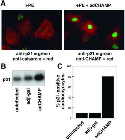
Up-regulation of p21CIP1 expression by CHAMP. (A) Primary neonatal rat cardiomyocytes were infected with adCHAMP or were uninfected and then incubated in serum-free medium containing PE (20 μg/ml) for 2 d. Uninfected cultures (Left) were stained with anti-calsarcin Ab to mark cardiomyocytes (red) and with anti-p21CIP1 Ab (green). adCHAMP-infected myocytes (Right) were stained with anti-CHAMP (red) and anti-p21CIP1 (green) Ab. The green p21CIP1 nuclear staining (Left) is from a fibroblast cell that does not have calsarcin staining. (B) p21CIP1 mRNA expression was measured by Northern blot analysis of RNA isolated from uninfected cultures and cultures infected with adβ-gal and adCHAMP. p21CIP1 mRNA expression was unaffected by adβ-gal, but was up-regulated by adCHAMP. (C) The percentage of cells in cardiomyocyte cultures that stained for p21CIP1 expression was determined. Weak p21CIP1 staining was seen in <10% of uninfected or adβ-gal-infected cultures. In contrast, >80% cells in adCHAMP-infected cultures showed strong p21CIP1 staining.

