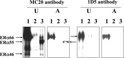
Figure 4.
Western blot analysis of ERα isoform abundance in the uteri (U) and the thoracic aorta (A) from ovariectomized ERα-WT (1), ERα-Δ2 KO (2), or ERα-Neo KO (3) mice. Antibody MC20 is directed toward the C-terminal domain and thus reveals all three isoforms (ERα66, ERα55, ERα46), and antibody 1D5 recognizes a sequence around the 119th amino acid of A/B domain and thus detects only ERα66. Each Western blot is representative of three separate experiments.

