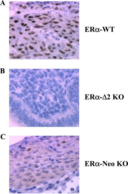
Figure 5.
Immunohistochemical analysis of ERα expression in the uterus from ERα-WT (A), ERα-Δ2 KO (B), and ERα-Neo KO mice (C). MC20 antibody was used to localize ERα in paraffin-embedded blocks of uteri from the three castrated strains to which E2 was given. Nuclei of uterus smooth muscle cells of ERα-WT were intensively stained. No immunoreactivity was detected in ERα-Δ2 KO mice. Nuclei and diffuse staining of the cytoplasm were observed in ERα-Neo KO mice.

