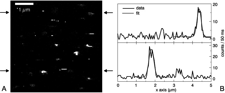
Figure 4.
(A) TPE fluorescence image of avidin proteins bound to latex spheres, each containing 340 Trp molecules (I = 750 kW/cm2 at 590 nm; integration time per pixel: 50 ms; image size: 5 × 5 μm). (B) Horizontal linescans at the positions indicated in A. Fitting the data to a Gaussian yields a focal spot size of ω0 = 350 nm.

