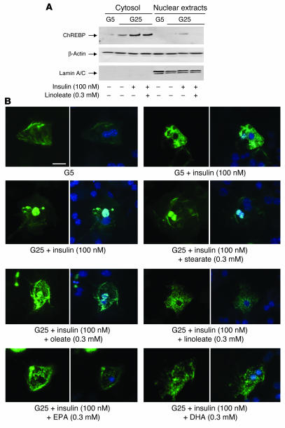
Figure 4.
PUFAs suppress nuclear translocation of ChREBP in cultured hepatocytes. After plating, hepatocytes were cultured for 24 hours in the presence of 5 mM glucose. Hepatocytes were then incubated for 24 hours in the presence of 5 or 25 mM glucose with or without 100 nM insulin and 100 nM dexamethasone containing or not 0.3 mM of albumin-bound linoleate. (A) Cytosolic and nuclear forms of ChREBP were measured. Representative Western blots of 4 independent cultures are shown. (B) Representative images of subcellular localization of GFP-fused ChREBP under 5 mM glucose with or without 100 nM insulin; and 25 mM glucose plus 100 nM insulin with or without 0.3 mM of albumin-bound stearate (C18), oleate [C18:1 (n-9)], linoleate [C18:2 (n-6)], EPA [C20:5 (n-3)], or DHA [C22:6 (n-3)]. For each condition, hepatocyte nuclei were specifically stained using DAPI (right panels). Scale bar, 10 μM.

