
Figure 1.
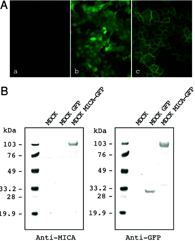
Expression of GFP-tagged MICA molecules in MDCK cells. (A) Green fluorescence signals were detected by using fluorescence microscopy in MDCK cells that were nontransfected (Left), stably transfected with the control GFP (Center), or MICA-GFP expression plasmids (Right). (B) Immunoblot analysis of GFP-tagged MICA molecules in MDCK cells: nontransfected MDCK cells (Control), MDCK cell lines stably expressing GFP (GFP), and MICA-GFP. Transferred membrane was stained with anti-MICA mouse serum (Left) or anti-GFP rabbit serum (Right). Protein size markers (kDa) are shown on the left.