
Figure 2.
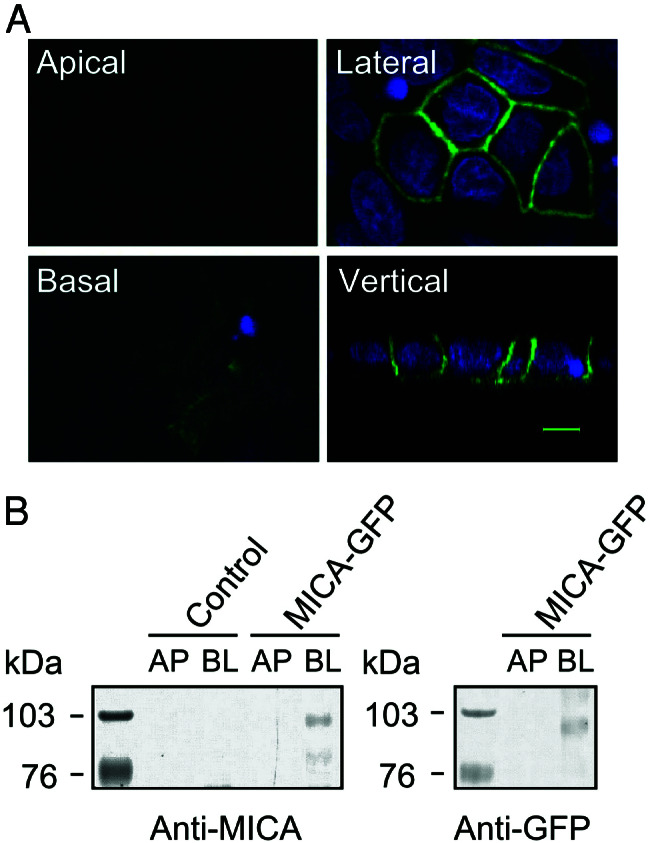
Intracellular localization of GFP-tagged MICA molecules in polarized MDCK cells. (A) MDCK cells stably expressing GFP-tagged MICA molecules. MICA-GFP fluorescence is colored by green, and nuclei are colored by blue. Fluorescence signals were analyzed by laser-scanning confocal microscopy. Confocal fluorescence micrographs (x–y plane) were acquired at the apical (Upper Left), lateral (Upper Right), and basal region (Lower Left). Confocal fluorescence micrographs (x–z plane) showing the distribution of MICA-GFP along the apical-basal axis (vertical). The scale bar is 5 μm. (B) Domain-selective cell-surface biotinylation assay of MDCK cells. Stably transfected MDCK cells expressing GFP-tagged MICA molecules were biotinylated from either the apical (AP) or basolateral (BL) domain. Biotinylated proteins were recovered by immunoprecipitation with anti-MICA mouse serum (Left) or anti-GFP rabbit serum (Right) and detected by SDS/PAGE-immunoblot analysis by using HRP-conjugated streptavidin. Control lanes on the left panel correspond to untransfected cells. Protein size markers (kDa) are shown on the left.