Fig. 3.
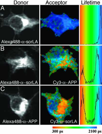
FLIM of APP695 and sorLA interaction in N2A cells. Shown are intensity images of sorLA (A and B) and APP695 (C) immunocytochemistry using donor fluorophore Alexa Fluor 488-conjugated antibodies (Left), pseudocolored FLIM images (Center), and lifetime histograms (Right), indicating shortening of lifetime from blue to orange/red in the absence (A) or presence (B and C) of acceptor fluorophore Cy3. (A) Cells stained for the amino-terminal domain of sorLA with donor fluorophore Alexa Fluor 488 in the absence of acceptor (lifetime of 2,007 ± 12 psec). (B) Cells stained for sorLA with donor fluorophore Alexa Fluor 488 in the presence of acceptor Cy3 on the ectodomain of APP695 (lifetime reduced to 1,365 ± 139 psec). (C) Cells stained for APP with donor Alexa Fluor 488 in the presence of acceptor Cy3 on sorLA (lifetime reduced to 1,481 ± 228 psec).

