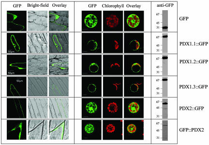
Fig. 3.
Subcellular localization of AtPDX1.1-1.3 and AtPDX2. (Left) Transient expression of the indicated GFP fusion proteins in onion epidermal cells. (Center) The transient expression observed in isolated A. thaliana protoplasts. (Scale bars: 10 μm, unless indicated otherwise.) (Right) Western blot of crude extracts is shown from isolated A. thaliana protoplasts expressing the indicated constructs after immunodecoration with anti-GFP, indicating that the fusion proteins were intact.

