
Figure 7.
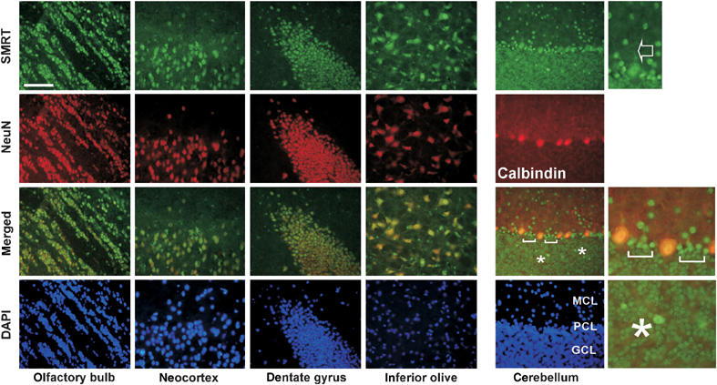
SMRT is enriched in neuronal cells in the mouse brain. Frozen brain sections prepared from 8-week-old wild-type (FVB/N) mice were co-immunostained with SMRT(972–1151) antibody (FITC, green) and NeuN or Calbindin antibody (Texas red, red). The staining of SMRT in the dendritic regions of Purkinje cells is indicated with an arrow. Brackets and asterisks mark elevated SMRT expression in the cells adjacent to Purkinje cells and within the granular cell layer, respectively. Scale bar: 1000 μM.