
Figure 7.
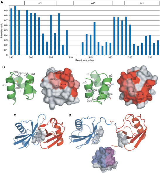
Molecular model for the interaction of Mud1 UBA with K48-linked Ub2. (A) Intensity changes by cross-saturation of the 15N–1H cross-peaks in 2H, 15N-Mud1 UBA in complex with unlabeled K48-Ub2. (B) Primary (left) and secondary (right) binding sites on Mud1UBA as identified by NMR cross-saturation. Resonances showing intensity ratios <0.5 or 0.3 are displayed on the molecular surface of Mud1 UBA in light or dark red, respectively. (C) Closed conformation of Ub2, based on the crystal structure obtained under basic conditions (PDB accession code 1AAR) (Cook et al, 1992). The hydrophobic patches on each Ub moiety interact with each other. The proximal and distal moieties of Ub2 are colored in red and blue, respectively. (D) Open conformation of Ub2, in equilibrium with closed conformation in solution. The two hydrophobic clusters formed by residues Leu8, Ile44, His68 and Val70 are available for binding of a single UBA domain via a primary (purple) and a secondary (blue) Ub-binding sites.