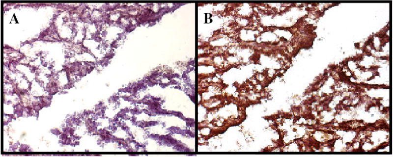
Fig. 3: Example - Immunohistochemical Analysis of Her-2/neu Expression in a Rat Mammary Tumor.

Paraffin embedded mammary tumor tissue taken from rat #538 was analyzed as described in Table 2 legend and the Methodology Section and presented here as an example. Brownish pigment is indicative of the presence of erbB-2/neu expression. A: IgG2a control antibody, B: Anti-rat Her-2/neu monoclonal antibody, 7.16.4.
