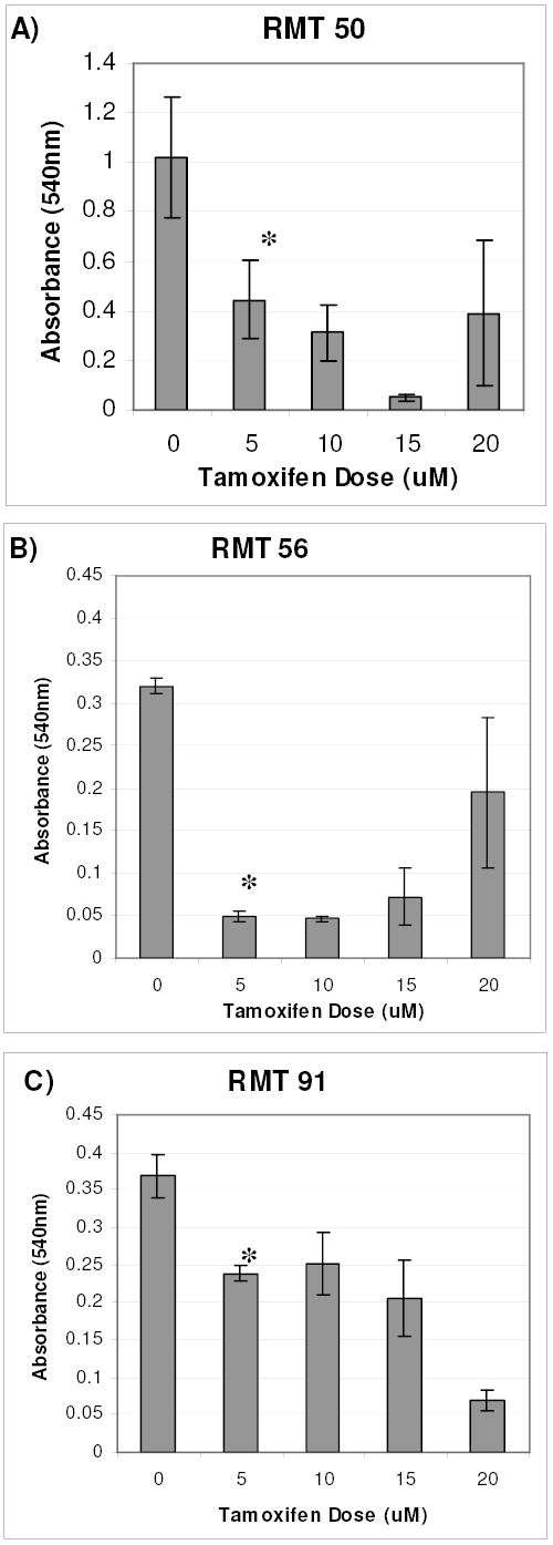
Fig. 6: In Vitro RMT Cell Growth Response to Tamoxifen.

RMTs 50, 56, and 91 were exposed to the indicated concentrations of tamoxifen for 5 days. The MTT assay was performed to determine relative numbers of viable cells in each group. Replicates of 5 were analyzed for each treatment group and the means +/− standard deviations are represented. The assay was repeated and similar results were obtained (data not shown). * mean absorbance (540nm) of 5 uM tamoxifen treated cells was significantly less than untreated cells. ANOVA (p<0.05), Fisher 95% Independent Confidence Interval.
