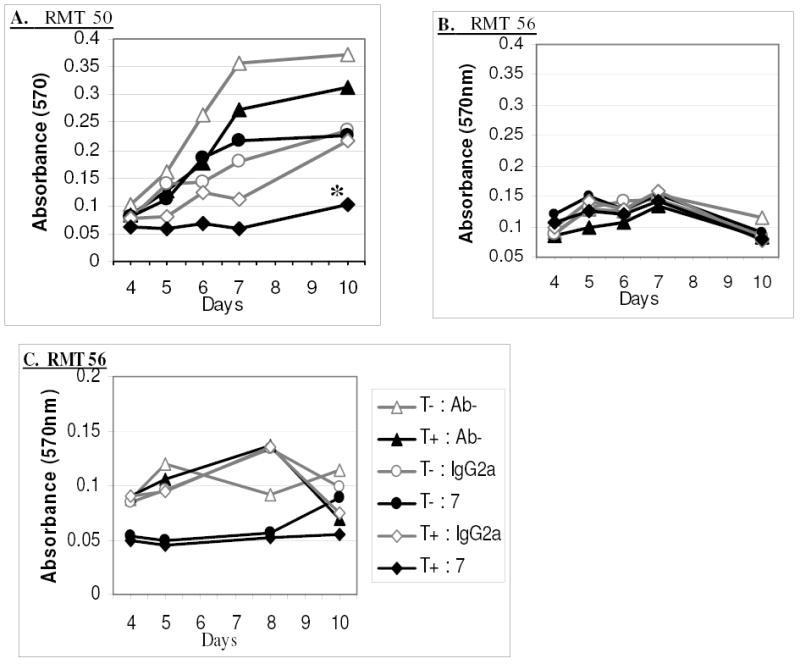
Fig. 9: In Vitro RMT Cell Growth Response to Combined Tamoxifen and Anti-Her-2/neu MAb 7.16.4 Treatment.

RMT 50 (A) and RMT 56 (B & C) were seeded in 5 replicate plates and incubated with 0.25 uM tamoxifen (T), 50 ug/ml MAb 7.16.4 (7), 50 ug/ml IgG2a, without antibody (Ab-), or any combination of these as indicated. C) 0.5 uM tamoxifen, 200 ug/ml MAb 7.16.4, and 200 ug/ml IgG2a were used. The MTT assay was performed on one of the plates at each of the specified days to determine the relative numbers of viable cells in each group. Replicates of 5 were analyzed for each treatment group on each day and the means are represented. * mean absorbance (540nm) was significantly less than other treatment groups. ANOVA (p<0.05), Tukey 95% Simultaneous Confidence Interval.
