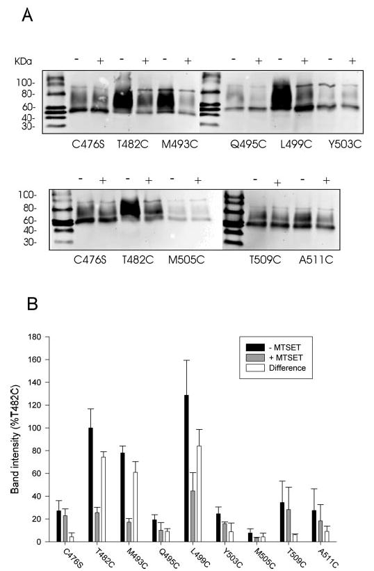
Fig. 8. Labeling of cysteine-substituted mutants with MTSEA-biotin.

A, HRPE cells expressing some of the cysteine mutants were first preincubated with sodium buffer with or without 1 mm MTSET to label extracellularly accessible cysteines. The preincubation solution was washed away, and the cell monolayers were biotinylated with MTSEA-biotin and transferred to Western blots as described under “Experimental Procedures.” The blots were treated with anti-NaDC-1 antibodies (1:1000 dilution). The negative control mutant, C476S, and the positive control, T482C, were included in each biotinylation experiment. +, pretreatment with MTSET; −, pretreatment with sodium buffer. The chemiluminescent size standards, Magic Mark Western Standards (Invitrogen), are shown, and mass (in kDa) is indicated to the left. B, summary of MTSEA biotinylation results. Western blots, such as those shown in A, were quantitated and expressed as a percentage of the T482C intensity from the same blot. The difference between the signal in the presence and absence of MTSET, which represents the MTSET-inhibitable binding of MTSEA-biotin, is also shown. Bars represent mean ± range (n = 2 blots).
