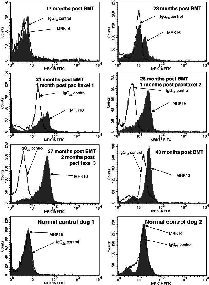
Figure 2.
Expression of P-gp in canine peripheral blood. For detection of P-gp, peripheral blood cells were stained with mAb MRK16 or a nonbinding isotype control, followed by staining with an FITC-labeled anti-mouse IgG antibody. Histograms display expression of P-gp in peripheral blood cells of animal M862 at indicated time points. As a negative control, blood cells from two normal, untreated dogs were analyzed (Bottom).

