
Figure 6.
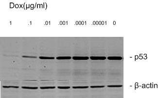
Expression of p53 protein by Dox controlled quantitatively. After p53-13 cells were cultured in 0 and 1, 0.1, 0.01, 0.001, 0.0001, 0.00001 μg/ml Dox for 24 h, expression of p53 protein was examined by Western blotting. β-actin was served as an internal control.