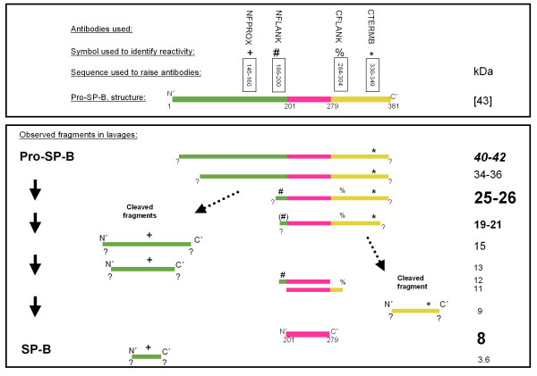
Figure 1.
Schematic diagram of pro-SP-B and its processing to SP-B. Upper panel: Indicated are the antibodies used, the symbols for their identification, the amino acid stretches against which the antibodies were developed, and a diagram of the structure of pro-SP-B. Lower panel: The molecular weight and the reactivity of the antibodies (in the absence, but not in the presence of the competing peptides) during Western blotting is indicated. The sizing of the letters used for indication of the molecular weights is proportional to the frequency at which the bands were observed (biggest: common >75% of subjects, 2nd biggest: frequent, in <75 but >50% of the subjects, 3rd biggest: sporadic, in <50 but >25% of the subjects, smallest: rare, in <25% of the subjects). The sequence of SP-B within the pro-SP-B sequence is indicated in pink. All bands were analyzed under reducing conditions.

