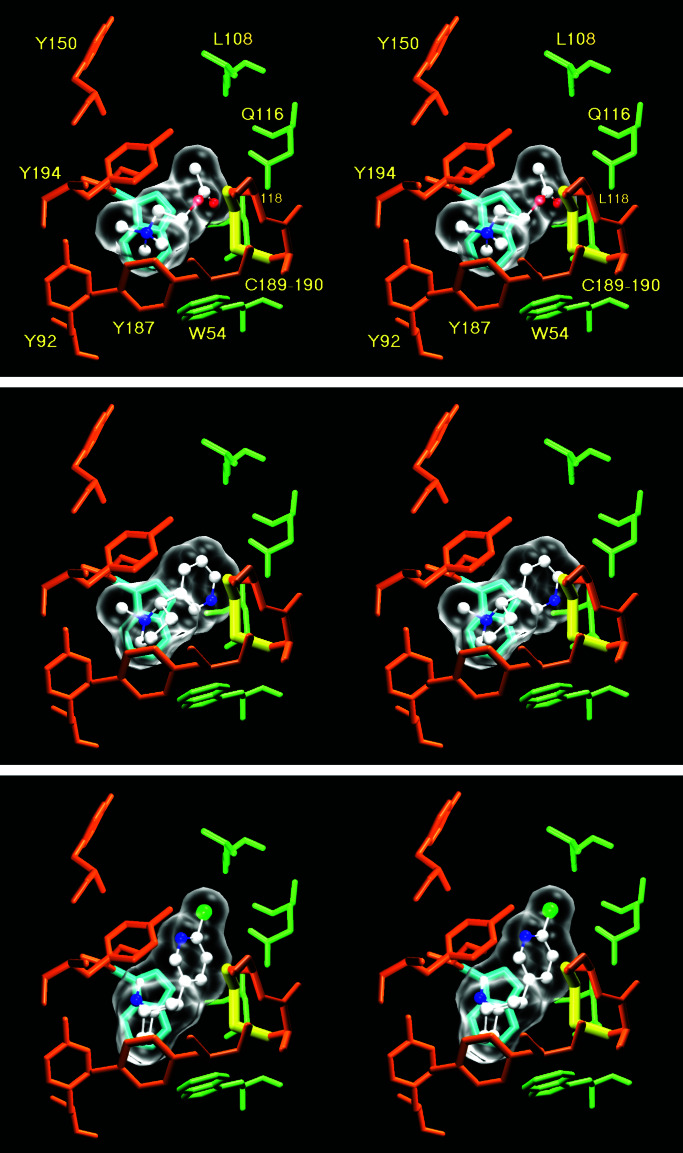
Figure 3.
Stereoscopic representation of the dockings of acetylcholine (Top), nicotine (Middle), and epibatidine (Bottom) onto the model of the amino-terminal part of α7. Oxygen atoms are red spheres, whereas nitrogens are blue, carbons are white, and the chlorine is green. The C189–190 disulfide bond is highlighted in yellow. W148, which makes a π-cation interaction with the ammonium, is highlighted in cyan. Note that all the sidechains are in the same position. Indeed, autodock does not allow sidechain movements during the docking process.

